Meltem Uçar1 ![]() ,
Orhan Deger2,
Asuman Yigit Gerigelmez2,
Sevil Cengiz3,
Yasam Barlak4,
Ercüment Ovalı5
,
Orhan Deger2,
Asuman Yigit Gerigelmez2,
Sevil Cengiz3,
Yasam Barlak4,
Ercüment Ovalı5
For correspondence:-
Received: 19 April 2016 Accepted: 15 October 2016 Published: 29 November 2016
Citation: Uçar M, Deger O, Gerigelmez AY, Cengiz S, Barlak Y, Ovalı E. Effect of Turkish pollen and propolis extracts on caspase-3 activity in myeloid cancer cell lines. Trop J Pharm Res 2016; 15(11):2445-2449 doi: 10.4314/tjpr.v15i11.20
© 2016 The authors.
This is an Open Access article that uses a funding model which does not charge readers or their institutions for access and distributed under the terms of the Creative Commons Attribution License (http://creativecommons.org/licenses/by/4.0) and the Budapest Open Access Initiative (http://www.budapestopenaccessinitiative.org/read), which permit unrestricted use, distribution, and reproduction in any medium, provided the original work is properly credited..
Purpose: To investigate the apoptosis-inducing capacity of dimethyl sulfoxide (DMSO) extracts of bee pollen and propolis in HL-60 Myeloid Cancer Cell Lines.
Methods: DMSO extracts of pollen and propolis were incubated separately with HL-60 cells, and caspase-3 activity evaluated. In order to determine the cell cycle characteristics of HL-60 cells with and without extracts of pollen and propolis, the cells were analysed using flow cytometry.
Results: The DMSO extract of propolis (0.5 mg/mL) increased apoptosis from undetectable levels to 60.1 %, while maintaining cell viability. The DMSO extract of pollen (2 mg/ml) increased apoptosis from undetectable levels to 52.2 % while decreasing cell viability by 62 %. Caspase-3 activity in HL-60 cells incubated with DMSO extracts of pollen and propolis were 3.6- to 12-fold higher than in controls.
Conclusion: Turkish pollen and propolis individually increase apoptosis and the activity of caspase-3 in HL-60 cells. This finding indicates that bee products may have beneficial effects in the treatment of cancer.
Introduction
Pollen is produced by bees after collecting millions of pollen and they use it as a food [1]. Propolis is collected from trees and some plants which have resin. Bees use propolis for repairing their hive, and also use it as antiseptic against invaders [2]. Bee pollen involves polyphenolic compounds, flavonoids, proteins, essential free amino acids for humans, carbohydrates, vitamins, minerals depending on the geographic area and climate from which they were collected [1,3]. Propolis generally is composed of 50 % resin and vegetable balsam, 30 % wax, 10 % essential and aromatic oils, 5 % pollen and 5 % various other substances, including organic debris. Hundreds of chemical compounds have been identified from propolis.
The main chemical classes present in propolis are flavonoids, phenolics, and various aromatic compounds but there is a standardization problem [2]. Bee pollen has anti-atherosclerotic, anti-neoplastic, antimicrobial, antimutagenic and antioxidant activities [4]. Propolis also has anti-bacterial, anti-viral, anti-fungal activities and immune activating and cytotoxic effect on cancer cells, anti-inflammatory, anaesthetic and antioxidant activities [2]. Antioxidant activities of both pollen and propolis originate from their polyphenolic compounds and flavonoids. Both pollen and propolis contain considerable amounts of poyphenolic substances such as quercetin, caffeic acid and caffeic acid phenethyl ester (CAPE), pinocembrin, galangin, etc. which may act as potent antioxidants [1,4,5].
Apoptosis is programmed cell death and occurs when the cell life cycle ends or various apoptosis triggers such as radiation, hazardous chemicals, drugs damage the cell [6,7]. Caspase-3 activation has an effector role in both the receptor and mitochondria mediated apoptosis [8]. Caspase cascade and cell death can be initiated by caspase-3 activation [9].
DMSO extracts of propolis and pollen at different concentrations were used to investigate anti-tumor and apoptosis-inducing activity in myeloid HL-60 cell line with lymphoid cell culture as a control. Flow-cytometry and spectrophotometric caspase-3 activity were used to determine the apoptosis-inducing activity.
Methods
Propolis and pollen
Propolis and pollen samples were obtained from honey-bees (Apis mellifera L.) in the region of Yomra, Trabzon, Turkey rich in Picea orientalis, Fagus orientalis, Castanea sativa, Rhodendron ponticum, Rhododendron luteum and Rubus caucasicus [10]. They were provided by Trabzon Agricultural Development Cooperative.
Preparation of DMSO extracts of pollen and propolis
Natural propolis samples were pulverized by grinding (Retsch, ZM 200). A 5 g pollen and ground propolis samples were dissolved in 5 mL of DMSO (100 % w/v) by continuous mixing for 5 h, then kept at 37 oC in water bath overnight. Extracts of 4 mL (1000 mg/mL) obtained by centrifuging at 800 g for 15 min were filtered and final volumes were made up to 10 mL with deionised water. Filtration procedure was repeated and the final extract was adjusted to 10 mL by deionised water to give a stock concentration of pollen and propolis extracts of 240 mg/mL. Working solutions at final concentrations of 0, 0.125, 0.25, 0.5, 1 and 2 mg/mL were prepared in phosphate buffered saline (PBS).
HL-60 myeloid cancer cell line and MNC cell isolation
HL-60 cell line (Hematology Laboratory, Faculty of Medicine, KTU, (American Type Culture Collection) was maintained as recommended by the manufacturer, while mononuclear cells (MNC) were obtained from 50 mL of heparinized peripheral blood supplied by the Blood Bank with Ficoll density gradient (density 1000 lymphoprep Nycomed Pharma, Oslo) and subsequent basic cell culture procedures using RPMI 1640 medium.
Cell culture and incubation of pollen and propolis extracts
MNC and HL-60 myeloid cancer cells were incubated in propolis and pollen extracts of final concentrations of 0, 0.125, 0.25, 0.5, 1 and 2 mg/mL in RPMI 1640 containing 10 % fetal calf serum, 1 % penicillin and streptomycin under 5 % CO2 pressure at 37 oC for 72 h.
Reagents
All reagents used were of analytical grade. DMSO was purchased from Sigma, Germany.
Cell viability
Pellets of cells were washed twice with PBS, then suspended in 25 µL PBS, then 1 µL of a mixture of fluorescent dyes containing 100 µg/mL acridine orange and 100 µg/mL ethidium bromide (AO/EB) was added to the cells and mixed gently. A drop of the mixture was placed on a microscope slide and covered with a coverslip. The cells were visualized under a fluorescent microscope, using excitation wavelength at 510 nm [11]. All experiments were done in duplicate.
Determination of apoptosis by flow-cytometric fluorescence analysis
The DNA content of the stained nuclei was analyzed by flow cytometry (Coulter Epics Elite ESP). The distribution of DNA content was expressed as G1, S, and G2/M phases. The cells with DNA content less than G1 were distributed in pre-G1 (hypodiploid cells) and expressed as the apoptotic phase [12]. All experiments were performed in duplicates.
In vitro caspase-3 assay
The activity of CPP32/caspase-3 was determined in cell extracts using a colorimetric protease assay kit (Biosource International USA, Catalog no: CPP32-KHZ 0022). The cells were collected and lysed with cell lysis buffer. Cell lysate was incubated with reaction buffer containing 5 µL of peptide substrate (DEVD-pNA) for 2 h at 37 oC. The cleavage of colorimetric peptide substrate was monitored by pNA (p-nitroanilide) liberation using a microtiter plate reader at 405 nm with Spectra Fluor Plus, Tecan A 5082 spectrophotometer. The activity was expressed as fold change of absorbance value over that at 0 concentration. All experiments were performed in duplicates.
Statistical analysis
Data are expressed as mean ± standard deviation (SD) for variables. Compatibility with normal distribution was determined using Kolmogorov-Smirnov test. One-way ANOVA was used to compare differences among the groups. Comparison of two groups was done by Student’s t-test.
Results
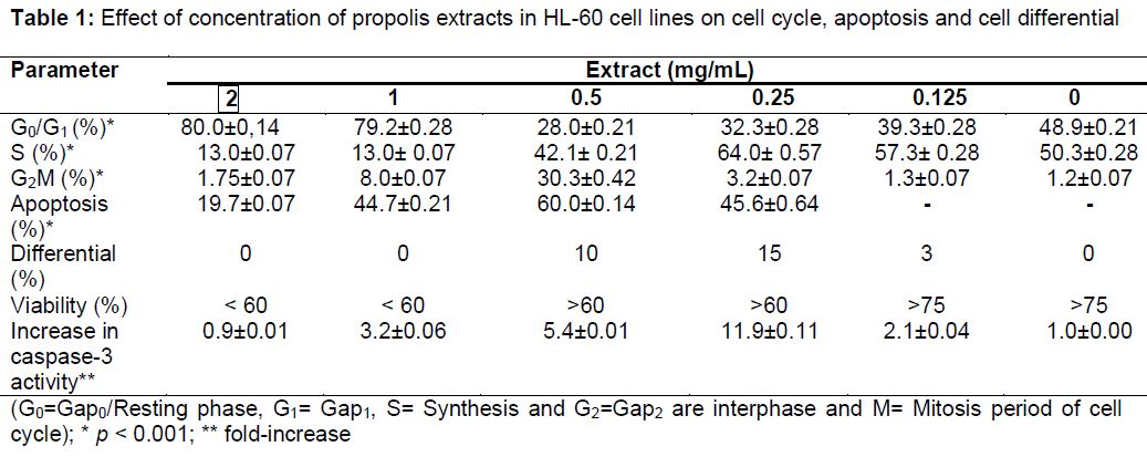
As shown in , significant differences were found among concentration groups. In particular, propolis extracts of 2 and 1 mg/mL had toxic effect on HL-60 cancer cells. Also, those concentrations did not lead to any differentiation and changed morphological appearances of the cells. However, 0.5, 0.25, 0.125 mg/mL propolis extracts increased HL-60 cancer cell number in the S phase and also increased mitotic activity. Cell viability was lower in cancer cells treated with 2 and 1 mg/mL propolis extracts (< 60 %). However, cell viability was higher (> 75 %) in cancer cells treated with 0.125 mg/mL propolis extracts. As shown in propolis extracts increased caspase-3 activity variously depending on concentration. Optimal increase occurred with 0.25 to 0.5 mg/ml extract. At these concentrations, apoptosis was high (45.1 to 60.1 %); differentiation was also high (10 to 15 %). However, the increase in caspase-3 activity in MNC cells was not as high.
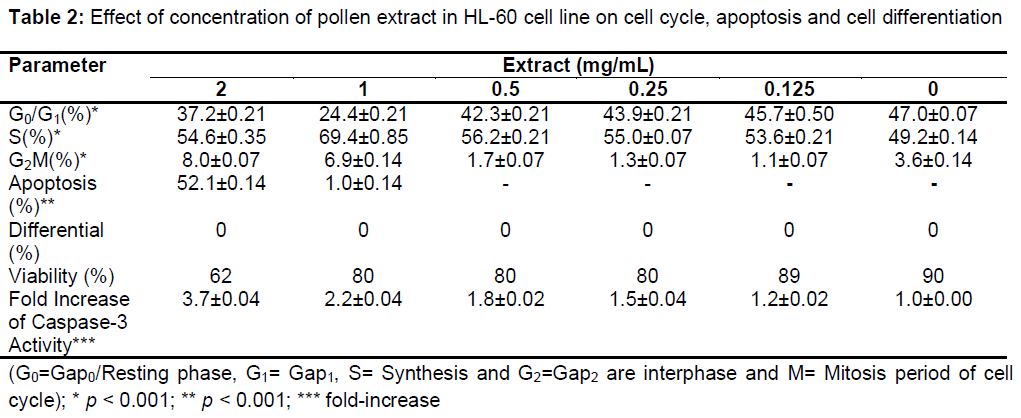
As shown in , significant differences were found among concentration groups. Pollen DMSO extract (2 mg/mL) had higher apoptotic activity (52.2 %) in HL-60 cancer cells than control and other groups. The highest concentration (2 mg/mL pollen DMSO extracts) also had increased cell number in which S and G2M phase were high. Caspase-3 activity also was higher at this concentration than others. Therefore, only pollen extract of 2 mg/mL seems to exert apoptotic activity. But, pollen extracts had no differentiation activity. In addition, DMSO pollen extract had a 1.75 fold increase in caspase-3 activity in MNC cells (data not shown).
Discussion
There are a lot of investigations about biological, biochemical, physiological, pharmaceutical and medicinal properties of polyphenolic compounds and flavonoids. There are also many studies about free radical scavenging activity and multiple biological activities of pollen and propolis including anti-inflammatory, immunomodulatory, anticarcinogenic and antioxidative or radical scavenging activity. All those studies suggested that pollen and propolis have these properties because of polyphenolic compounds and flavonoids they contain. Analysis of phenolic compounds and flavonoids of Turkish propolis was previously carried out in our laboratory. According to HPLC analysis, the main flavonoids found include galangin, naringenin, chrysin, kaempferol, quercetin and cinnamic acid derivatives for DMSO extracts and, caffeic and caffeoyl quinic acids for water extracts [13].
The main finding in the present study was that DMSO extracts of pollen and propolis at different concentrations promoted apoptosis in HL-60 cells by activating caspase-3. DMSO extract of propolis (0.5 mg/mL) increased apoptosis from undetectable to 60.1 %, while maintaining cell viability. But 1 and 2 mg/mL concentration of DMSO extract of propolis had cytotoxic effect on HL-60 cells and decreased cell viability to < 60 percent. However, 0.5, 0.25 and 0.125 mg/mL concentration of DMSO extract of propolis caused differentiation in HL-60 cells. On the other hand all concentrations of DMSO extracts of propolis increased caspase-3 activity in HL-60 cells when compared with control that is 0 mg/mL concentration. Fold-increase of caspase-3 activity was changed from 0.88 to 12.00 in HL-60 cells. Furthermore, only 2 and 1 mg/ml concentration of DMSO extract of pollen induced apoptosis in HL-60 cells from non-detectable level to 52.2 and 0.9 % respectively. Caspase-3 activity in cells which were incubated with DMSO extracts of pollen was found to be concentration dependent. It was changed from 1.25- to 3.65-fold.
Many chemopreventive agents act through the induction of apoptosis as a mechanism of antitumorigenesis. Eroğlu et al [14] and Gunduz et al [15] suggested that Turkish propolis can decrease cell division in tissue cultures of bladder cancer and may show antitumoral and apoptotic effect on leukemia cells. Chen et al [16] investigated the mechanism of CAPE-induced apoptosis in human leukemic HL-60 cells. It was found that CAPE entered HL-60 cells very quickly and inhibited their survival depending on the concentration and time. They also observed DNA fragmentation and morphological changes typical of apoptosis in the cells. CAPE caused rapid activation of caspase-3, down-regulation of Bcl-2 expression and up-regulation of Bax expression in HL-60 cells [16].
Jin et al [17] investigated the mechanism of CAPE-induced apoptosis in U937 cells. They showed that CAPE decreased cell viability in dose-dependent and time dependent manner, DNA fragmentation and nuclear condensation in cancer cells and apoptotic action accompanied by release of cytochrome c, reduction of Bcl-2 expression, increase of Bax expression [17].
Woo et al [18] also used a different component of propolis, chrysin in U937 cells and they showed that chrysin activated caspase-3 and inactivated Akt [18]. Propolin A, propolin B and propolin C, components of propolis, were used by Chen et al [19,20] in human melanoma A2058, HL-60, MCF-7, human neuroblastoma IMR-32, C6, murine melanoma B16F10, Hep.G2, human hepatocellular carcinoma Hep.3B and HT-29 cells. They showed that propolin A, propolin B and propolin C are cytotoxic to cancer cells. They also observed that those components induce apoptosis by decreasing procaspase-8, Bid, procaspase-3, DFF45 and PARP(poly ADP ribose polymerase) depending on the time and dose [19,20].
In the present study, propolis or pollen extracts alone were preferred to their polyphenolic or flavonoid components such as CAPE, galangin, pinocembrin etc. We suggest that those extracts may exert their anti-tumoral activities by boosting the immune system. Also Premratanachai and Chanchao [21] suggested that the mechanism of action of bee products in inhibiting tumor growth in vitro and in vivo is mediated via apoptosis, necrosis and lysis of tumor cells [21].
Conclusion
Both DMSO extracts of Turkish propolis and pollen may induce apoptosis in high concentrations in cancer cells. Further studies are required to elucidate the mechanism of action in higher concentrations.
Declarations
Acknowledgement
References
Archives


News Updates